Pune
08048051757
+919834023072

Services

6814772a5d263414004dd4a4 Card 2

product image
Ultrasonography 3D/4D

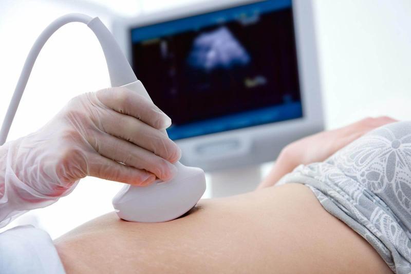
3D and 4D ultrasound tests provide enhanced visualization of the fetus during pregnancy, offering more detailed images and even real-time motion compared to traditional 2D scans. 3D ultrasound creates static, three-dimensional images, while 4D ultrasound adds the element of time, allowing for real-time viewing of the fetus's movements. 3D Ultrasound: Static 3D Images: Creates still, three-dimensional pictures of the fetus, allowing for a more detailed view of facial features and body structures. Improved Visualization: Provides a more comprehensive view of the fetus's anatomy compared to 2D ultrasound. Examples: Can be used to assess facial features like the nose and lips, and to examine the development of limbs and other body parts. 4D Ultrasound: Real-time Video: Adds the dimension of time to 3D imaging, creating a live video of the fetus's movements. Dynamic View: Allows observation of fetal actions like yawning, sucking, and limb movements in real-time. Enhanced Bonding: Provides a memorable experience for parents by allowing them to see their baby's actions as they happen. Key Differences: 3D: Produces still 3D images. 4D: Produces moving 3D images (real-time video). Benefits of 3D/4D Ultrasound: Early Detection: Can help detect certain fetal abnormalities like cleft lip, spinal problems, and other congenital disabilities. Improved Monitoring: Helps monitor fetal growth, amniotic fluid levels, and other aspects of pregnancy. Enhanced Parental Bonding: Provides a more engaging and intimate experience for parents to connect with their unborn child. Better Visualization of Fetal Heart: Can offer a clearer view of the heart structures, aiding in the diagnosis of heart defects. Safety Considerations: Generally Safe: Ultrasounds are considered safe for both mother and baby when performed by trained professionals.

6814772a5d263414004dd4a4 Card 2

product image
Digital X-Ray / Portable X-Ray (Home Visit)

Digital and portable X-ray services are now available for home visits in Pune, offering convenient diagnostic imaging for patients who may have difficulty traveling to a clinic or hospital. These services utilize portable X-ray machines to capture images at the patient's location, providing quick and effective assessments, especially for those with mobility issues. Key Features and Benefits: Accessibility: Portable X-ray services make it easier for individuals with mobility limitations, chronic illnesses, or those in remote locations to access diagnostic imaging. Convenience: Patients can undergo X-ray examinations in the comfort and convenience of their own homes, eliminating the need for travel. Efficiency: Portable X-ray machines offer quick results, allowing for timely diagnosis and treatment decisions. Digital Technology: Many portable X-ray units utilize digital imaging technology, which provides high-quality images and allows for immediate viewing and sharing of results. Wide Range of Applications: Portable X-rays can be used to assess various conditions, including bone fractures, lung issues, and other injuries or ailments. Finding Portable X-ray Services in Pashan Pune

6814772a5d263414004dd4a4 Card 2

product image
Fetal Ultrasound

A fetal ultrasound, also known as a sonogram, is a safe and common medical test that uses sound waves to create images of a baby developing inside the womb. It's a standard part of prenatal care, allowing healthcare providers to monitor fetal growth, development, and overall health, as well as assess amniotic fluid levels and placental position. Here's a more detailed explanation: Purpose: Fetal ultrasounds are used to: Confirm pregnancy and determine gestational age. Monitor fetal growth and development. Check for multiple pregnancies. Assess the location of the placenta. Evaluate amniotic fluid levels. Detect potential problems, like structural abnormalities or other medical issues. Procedure: The test is usually performed by placing a transducer (a device that emits sound waves) on the mother's abdomen or, in some cases, internally through the vagina. The sound waves bounce off the fetus and are processed into images. Safety: Ultrasound is considered a safe imaging technique as it doesn't use ionizing radiation like X-rays. Timing: Ultrasounds can be performed at various points during pregnancy, with the most common being a mid-trimester scan (around 18-22 weeks) for anatomical evaluation. What it can detect: Ultrasounds can detect various conditions, including: Structural abnormalities (e.g., anencephaly, spina bifida, cleft lip, heart defects). Down syndrome (trisomy 21) and other chromosomal abnormalities. Fetal growth restriction. Placental problems. Amniotic fluid abnormalities. Importance:

6814772a5d263414004dd4a4 Card 2

product image
Color Doppler

A Color Doppler test is a non-invasive ultrasound technique used to visualize blood flow in vessels and organs. It uses sound waves to create color-coded images that show the speed and direction of blood flow, helping to diagnose various vascular conditions and heart diseases. The test is painless, doesn't involve radiation, and is generally quick, taking around 10-30 minutes. What it is: A Color Doppler test is a specialized type of ultrasound that adds color to the images to represent blood flow. It uses high-frequency sound waves to create images of blood vessels and the blood flowing through them. The colors indicate the direction and velocity of blood flow, unlike standard ultrasound which provides static, black and white images. How it works: A transducer emits sound waves that bounce off blood cells. The echoes are captured and analyzed by a computer, which then converts them into color-coded images. The colors represent the speed and direction of blood flow. What it's used for: Diagnosing vascular conditions: Blood clots, narrowed arteries, and other blood flow problems. Assessing heart valve function: Evaluating blood flow through heart valves. Monitoring blood flow during pregnancy: Checking blood flow to the fetus, placenta, and umbilical cord. Detecting abnormalities in organs: Assessing blood flow in organs like the liver and kidneys. How to prepare for the test: You may be asked to wear loose-fitting clothes. For abdominal or pelvic scans, you may need to fast for a specific period (e.g., 6-12 hours). You may need to remove jewelry or other items that could interfere with the scan. During the test: A gel is applied to the skin to help the transducer glide smoothly. The transducer is moved over the area of interest. You may hear a whooshing sound as the machine processes the echoes. The test is generally painless, though you may feel some pressure from the transducer. Interpreting the results: The images are reviewed by a healthcare professional (doctor or radiologist). Abnormalities in blood flow patterns may indicate various conditions. The results are used to determine a treatment plan if needed. Is it safe? Yes, Color Doppler is a safe and painless procedure. It does not use radiation.

6814772a5d263414004dd4a4 Card 2

product image
ECG

An electrocardiogram (ECG or EKG) is a non-invasive test that measures and records the electrical activity of the heart. It's a quick and painless way to assess heart rhythm and rate, helping to diagnose various heart conditions. Here's a more detailed explanation: What it does: An ECG machine records the electrical signals produced by the heart's activity and displays them as waves on a graph or computer screen. How it works: Electrodes (small, sticky patches) are attached to the chest, arms, and legs. These electrodes are connected to the ECG machine, which detects and records the heart's electrical signals. Why it's done: ECGs can help diagnose heart attacks, irregular heartbeats (arrhythmias), and other heart problems. They can also assess the overall health of the heart, monitor the effectiveness of treatments, and evaluate how well implanted devices like pacemakers are working. When it's used: ECGs are used in various settings, including hospitals, clinics, and ambulances. Doctors may order an ECG if a patient experiences symptoms like chest pain, shortness of breath, dizziness, or palpitations. Types of ECGs: There are different types of ECGs, including resting ECGs (performed while lying down), stress or exercise ECGs (performed during physical activity), and ambulatory ECGs (monitored over a longer period, often at home).

6814772a5d263414004dd4a4 Card 2

product image
All Pathology Tests

Pathology tests encompass a wide range of examinations performed on bodily fluids, tissues, and even whole bodies to diagnose and monitor diseases. These tests can include blood tests, urine tests, stool tests, biopsies, and various imaging techniques. Specific examples include complete blood counts, lipid panels, liver function tests, and urinalysis. Types of Pathology Tests: Blood Tests: These are crucial for assessing overall health and identifying various conditions. Examples include complete blood counts (CBC), lipid panels (cholesterol and triglycerides), fasting glucose tests (for diabetes), thyroid function tests, and kidney function tests. Urine Tests: Urine tests, including dipstick and microscopy, can help detect kidney problems and other health issues. Stool Tests: These tests can help diagnose gastrointestinal issues and infections. Biopsies: A biopsy involves taking a tissue sample for microscopic examination to diagnose cancer and other diseases. Imaging Tests: Various imaging techniques, such as X-rays, ultrasounds, and MRIs, can be used in pathology to visualize internal structures and identify abnormalities. Genetic Tests: These tests analyze DNA to identify genetic predispositions to certain diseases. Common Pathology Tests: Complete Blood Count (CBC): Assesses red and white blood cell counts, hemoglobin levels, and other blood components to detect anemia, infections, and other conditions. Liver Function Tests (LFTs): Evaluate liver enzymes and proteins to assess liver health and diagnose conditions like hepatitis. Lipid Panel: Measures cholesterol and triglyceride levels to assess cardiovascular disease risk. Urinalysis: Examines urine for abnormalities to detect kidney and urinary tract issues. Thyroid Function Tests (TFTs): Measures thyroid hormone levels to diagnose thyroid disorders. Pap Smear: A cytology test to screen for cervical cancer. Cultures: Used to identify infectious agents like bacteria or fungi. Other Pathology Tests: Chemical Pathology: Analyzes chemical components in body fluids. Haematology: Studies blood and blood-forming tissues. Anatomical Pathology: Examines tissues and organs to diagnose diseases. Cytopathology: Studies cells for diagnostic purposes. Medical Microbiology: Identifies infectious agents. Immunopathology: Studies the immune system and its role in disease. Genetic Pathology: Studies the role of genes in disease. Forensic Pathology: Applies pathology principles to legal investigations. General Pathology: A broad term encompassing various aspects of pathology. Clinical Pathology: Focuses on laboratory testing of bodily fluids for disease diagnosis.

Phone Number

08048051757

Please keep 0 before dialling the number.

Mon-Thu: 10 AM - 2 PM • Fri: 3 PM - 7AM

Other Website Visit our other website
Address Office No: 8, Ground Floor, Precise Diagnostics, Vastu Nirvana, Baner - Pashan Link Rd, Pashan, Pune, Maharashtra 411021

Pune, India, 411021