
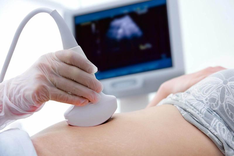
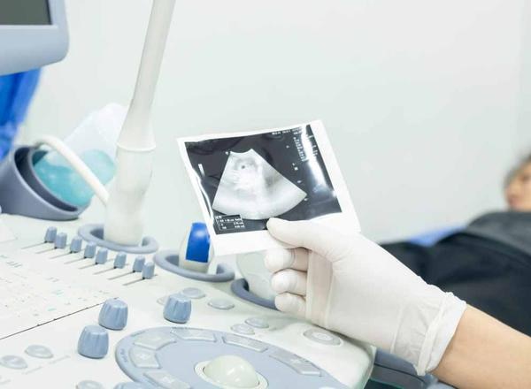
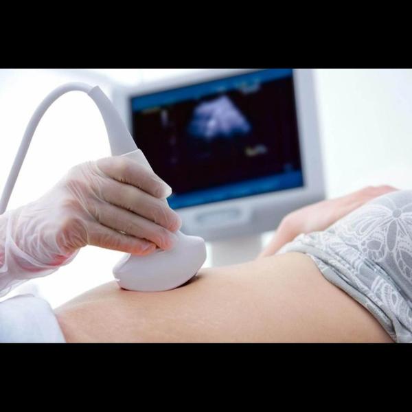
3D and 4D ultrasound tests provide enhanced visualization of the fetus during pregnancy, offering more detailed images and even real-time motion compared to traditional 2D scans. 3D ultrasound creates static, three-dimensional images, while 4D ultrasound adds the element of time, allowing for real-time viewing of the fetus's movements. 3D Ultrasound: Static 3D Images: Creates still, three-dimensional pictures of the fetus, allowing for a more detailed view of facial features and body structures. Improved Visualization: Provides a more comprehensive view of the fetus's anatomy compared to 2D ultrasound. Examples: Can be used to assess facial features like the nose and lips, and to examine the development of limbs and other body parts. 4D Ultrasound: Real-time Video: Adds the dimension of time to 3D imaging, creating a live video of the fetus's movements. Dynamic View: Allows observation of fetal actions like yawning, sucking, and limb movements in real-time. Enhanced Bonding: Provides a memorable experience for parents by allowing them to see their baby's actions as they happen. Key Differences: 3D: Produces still 3D images. 4D: Produces moving 3D images (real-time video). Benefits of 3D/4D Ultrasound: Early Detection: Can help detect certain fetal abnormalities like cleft lip, spinal problems, and other congenital disabilities. Improved Monitoring: Helps monitor fetal growth, amniotic fluid levels, and other aspects of pregnancy. Enhanced Parental Bonding: Provides a more engaging and intimate experience for parents to connect with their unborn child. Better Visualization of Fetal Heart: Can offer a clearer view of the heart structures, aiding in the diagnosis of heart defects. Safety Considerations: Generally Safe: Ultrasounds are considered safe for both mother and baby when performed by trained professionals.

Pune
08048051757
+919834023072
 Chat with us

Precise Diagnostics
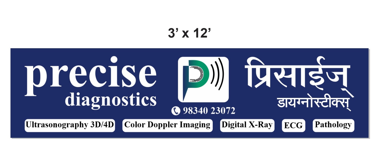
Precise Diagnostics
About Precise Diagnostics
The Best Diagnostic Center in Pune
Services
Featured ServicesWhat We Do


Have any custom requirements?
Updates
Read What’s Latest

Testimonials
View All
Taj Hmar
Amazing experience. Doctor is kind, humble and gives you priority. Doctor gave detailed information of my medical report...
Read MoreAkshay Kadam

Good experience.. I went there for USG abdomen & pelvis.. the doctor was very good and was able to answer all the questi...
Read MoreShubham Gavali

First visit itself was very nice. Staff is well mannered. Doctor is experienced. Thank you for the excellent service 👍�...
Read More
×
![]()